
"The Scan Shows a Swollen Kidney" - A Parent's Guide to Antenatal Hydronephrosis

Published 25 Sept 2026 · Updated 25 Sept 2026 · Medically reviewed 25 Sept 2026 · 10 min read
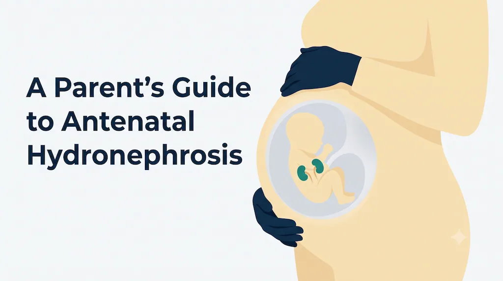
Your pregnancy scan report mentions hydronephrosis, pyelectasis or an "APD" measurement, and you're worried about your baby's kidney. This guide explains what the finding means, why most babies do well, and which steps after birth determine whether your baby needs any treatment at all.
In this article
What is antenatal hydronephrosis?
Hydronephrosis means that the part of the kidney that collects urine, called the renal pelvis, is wider than usual. Antenatal means it was seen before birth, on a pregnancy ultrasound.
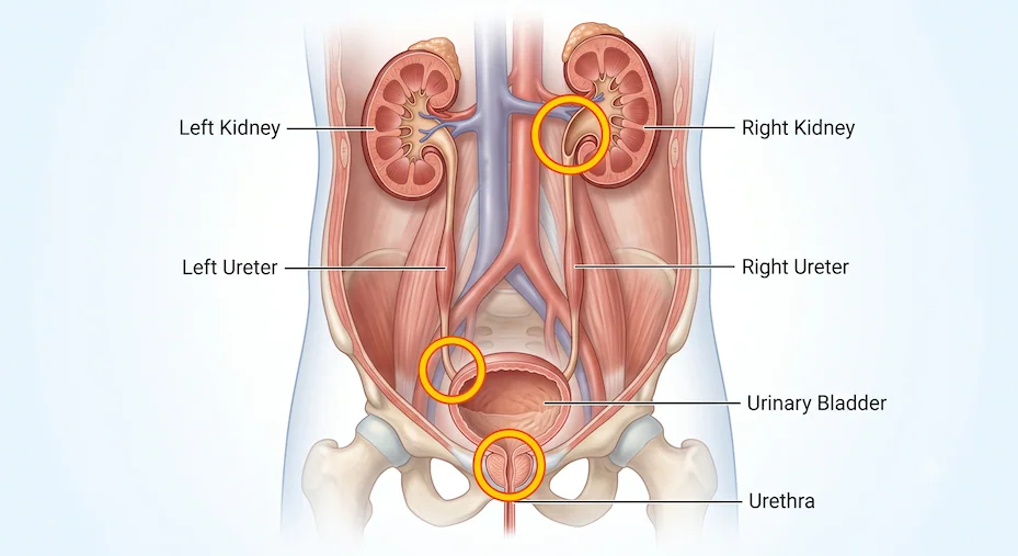
Your baby's kidneys start making urine early in pregnancy. The urine collects in the renal pelvis, drains down a thin tube called the ureter into the bladder, and passes out into the amniotic fluid. If urine drains a little slowly, or if the system is simply more stretchy than average, the renal pelvis looks wider on the scan.
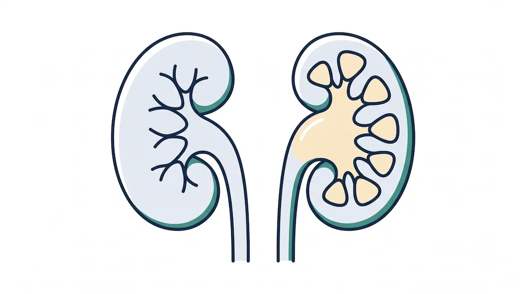
This is one of the most common findings on pregnancy ultrasound, seen in roughly 1 to 2 of every 100 pregnancies. Seeing it does not mean something is wrong. It means your baby's kidneys need to be followed up.
What does "APD" on the report mean? APD (anteroposterior diameter) is the width of the renal pelvis measured from front to back, in millimetres. It is the number most doctors use to judge how significant the dilation is.
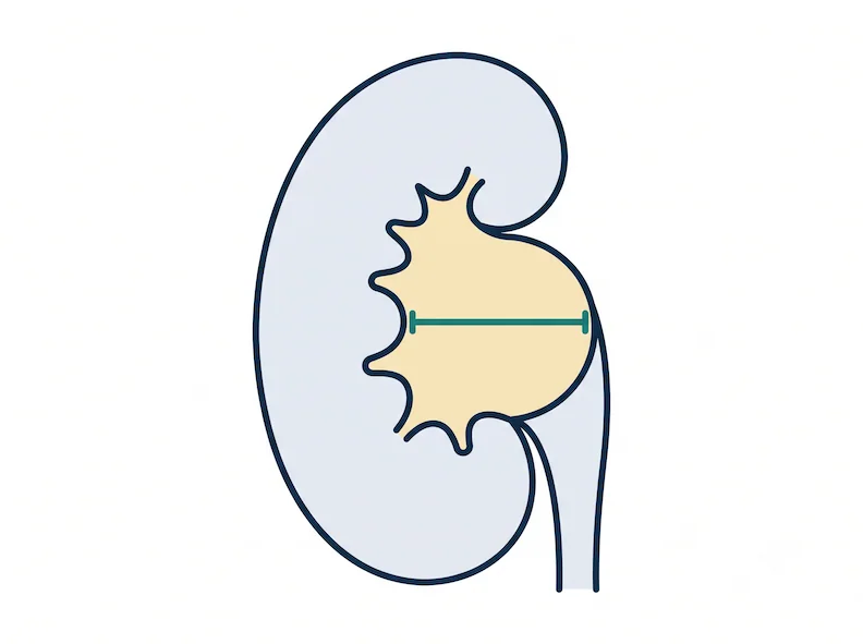
APD: a general guide
- 16–27 weeks: under 4 mm is usually normal. 4 mm or more needs follow-up.
- 28 weeks onward: under 7 mm is usually normal. 7 mm or more needs follow-up.
- A single reading varies with the baby's position and bladder filling. The trend across scans matters more than any one number.
What causes it?
In most babies, no blockage is ever found. The drainage system is simply wider than average during development and settles over time. When there is an underlying cause, it is usually one of the following:
- Transient dilation, the most common: the system looks wide but drains normally, and it often improves before or after birth without any treatment.
- PUJ obstruction: a narrowing where the renal pelvis joins the ureter slows drainage. It is the most common cause that sometimes needs surgery.
- Vesicoureteric reflux (VUR): urine flows backward from the bladder toward the kidney, which can raise the risk of urine infections.
- Megaureter: the ureter itself is wide, often because of a narrowing near the bladder.
- Posterior urethral valves (boys only): a blockage in the urine passage below the bladder. It usually affects both kidneys, and it is important to identify early.
- Duplex kidney or ureterocele: the kidney has two drainage systems, and one of them may be blocked.
How is it detected ?
Parents do not notice antenatal hydronephrosis. It is picked up on a routine pregnancy ultrasound, most often on the anomaly scan at 18–22 weeks or on a later growth scan. The report may use terms such as hydronephrosis, pyelectasis, pelviectasis, renal pelvic dilatation, UTD or APD. These all describe the same basic finding.
After birth, most babies with mild dilation look and behave completely normally. That is why planned follow-up scans matter: they find the few babies who need attention before any symptoms appear.
Your doctor will also look beyond the APD number: whether one or both kidneys are affected, whether the smaller branches (calyces) or the ureter are dilated, how the kidney tissue looks, whether the bladder is normal, and whether the amniotic fluid level is normal.
Findings that make the picture more significant than the APD alone:
- Both kidneys affected
- Dilated ureter
- Thick-walled or enlarged bladder
- Reduced amniotic fluid
- Abnormal-looking kidney tissue
When should you see a paediatric surgeon?
During pregnancy. For most mothers, nothing changes: the timing of delivery, the mode of delivery and routine care stay the same, and hydronephrosis alone is not a reason for early delivery. A consultation with a paediatric surgeon or paediatric urologist before delivery is useful when the dilation is significant, involves both kidneys, or comes with other findings such as a dilated ureter, an abnormal bladder or low amniotic fluid.
In these situations, it helps to plan delivery at a hospital with a NICU and a paediatric surgical team, and to meet that team in advance so everyone knows what will happen in the first days after birth.
After birth. Your baby should be reviewed by a paediatric surgeon or urologist if the postnatal scan shows significant or worsening dilation, a dilated ureter, a duplex kidney, or any bladder abnormality, or if your baby has a urine infection.
How is it diagnosed?
The pregnancy scan raises the question. Tests after birth answer it.
1. First ultrasound after birth. This is usually done after the first 48 hours of life, typically within the first week or two. In the first two days a baby makes relatively little urine, so a scan done too early can make the kidneys look falsely normal. When both kidneys are severely affected, a blockage below the bladder is suspected, or the baby has only one kidney, the scan is done earlier, often before discharge from the hospital.
2. Repeat ultrasound at around 4–6 weeks shows whether the dilation is improving, stable or worsening.
3. Further tests, only if needed. Most babies with mild dilation need only ultrasound follow-up. Some may need:
- MCU (micturating cystourethrogram): Dye is placed in the bladder through a fine tube, and X-ray images are taken while the baby passes urine. It checks for reflux and for blockages below the bladder.
- Diuretic renogram (DTPA or EC scan): A small tracer injection shows how well each kidney works and how well it drains. It is usually done after 6–8 weeks of age, when the kidneys have matured enough for reliable results.
Doctors often grade the findings using the UTD (Urinary Tract Dilation) or SFU system. The grade, together with how it changes over time, guides whether your baby needs only scans, further tests, or treatment.
Treatment and the right age for it
Most babies with antenatal hydronephrosis do not need surgery. Many mild cases improve or resolve on their own over months. For these babies, "treatment" means planned ultrasound follow-up.
Preventive antibiotics. Some babies, particularly those with a dilated ureter, suspected reflux or significant dilation, may be advised a small daily dose of antibiotic to protect against urine infection while tests are completed. Many babies do not need this. The decision is individual.
When surgery is considered. An operation is considered when follow-up shows reduced or falling kidney function, dilation that keeps increasing, repeated urine infections or other symptoms, or a blockage that will not resolve on its own.
The right timing depends on the cause. Posterior urethral valves are treated soon after birth, once the baby is stable, because the blockage affects both kidneys and the bladder. PUJ obstruction is usually operated on only when follow-up shows that the kidney needs it. This may be in the first months of life or later, which is why regular follow-up matters more than any fixed age.
The most common operation, for PUJ obstruction, is pyeloplasty. The narrowed segment is removed, and the renal pelvis is rejoined to the ureter so urine can drain freely. Depending on the child's age and anatomy, it can be done as an open operation or by keyhole (laparoscopic) surgery.
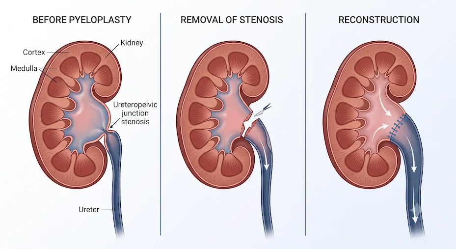
The goal of every step, from scans and tests to antibiotics and surgery, is the same: to protect your baby's kidney function over the long term, and to intervene only when the kidney needs it.
Caring for your baby at home and after treatment
During follow-up (most babies). Feed, bathe, vaccinate and care for your baby as usual. No special diet or restriction is needed. Keep all scan reports and CDs together in one folder, and bring them to every visit so the trend can be compared.
If your baby is on preventive antibiotics, give the dose once daily at the same time, and do not stop it without discussing it with your doctor. Tell any doctor treating your baby for another illness that your baby is on this medicine.
Watch the nappies. A wet nappy several times a day is reassuring. In boys, notice whether the urine comes as a good stream. Keep the nappy area clean, and change nappies promptly.
If your baby has had surgery, most babies are feeding within a day and go home after a short hospital stay. Your surgeon will explain whether a small internal tube (stent) has been placed to support healing, and when it will be removed. Follow-up scans after surgery check that drainage has improved.
The bottom line A swollen kidney on a pregnancy scan is a finding to follow up, not a diagnosis to fear. With timely scans after birth, further tests only when needed, and treatment only when the kidney actually needs it, most babies grow up with healthy kidneys. Book a consultation to go through your scan reports and plan your baby's follow-up.
Frequently asked questions
References
- Nguyen HT, Benson CB, Bromley B, et al. Multidisciplinary consensus on the classification of prenatal and postnatal urinary tract dilation (UTD classification system). J Pediatr Urol. 2014;10(6):982–998.
- Nguyen HT, Herndon CDA, Cooper C, et al. The Society for Fetal Urology consensus statement on the evaluation and management of antenatal hydronephrosis. J Pediatr Urol. 2010;6(3):212–231.
- EAU/ESPU Guidelines on Paediatric Urology, current edition: dilatation of the upper urinary tract.
Have a question about your child?
This article is for general education and does not replace a clinical examination. Every child's condition is different — please consult a qualified paediatric surgeon for advice specific to your child.
Helpful next reads for parents.
Parent Guide
PUJO & PUV in Children: Silent Causes of Kidney Damage Parents Should Know
A parent-friendly guide to PUJO and PUV, from antenatal hydronephrosis to the tests and treatments that help protect long-term kidney function.
Read related guideParent Guide
Recurrent Vomiting in Children: When Is It a Surgical Problem?
A practical guide for parents on repeated vomiting in children, warning signs of surgical causes, and when urgent evaluation is needed.
Read related guideParent Guide
Lumps and Swellings in Children: Which Ones Need Surgery?
A clear guide for parents to common lumps and swellings in children, which ones are usually harmless, and which need surgical review.
Read related guide